
The human body is so complex. The way all the parts work together to lead to a functioning whole is something that scientists may never fully understand. However, what we have managed to learn about human anatomy is quite fascinating!
Take the nervous system, for example. It’s basically the command center of the entire body, controlling everything you think and do. It consists of the central nervous system, comprised of your brain and spinal cord, and your peripheral nervous system, which includes a variety of nerves that extend to the rest of your body.
Of these nerves, some are larger than others. So which one is the largest cranial nerve? Keep reading to find out!
Structure and Function of a Nerve Cell
Nerve cells make up the brain and nervous system and interact with one another to allow for sensory and motor function.
©iStock.com/Vitalii Dumma
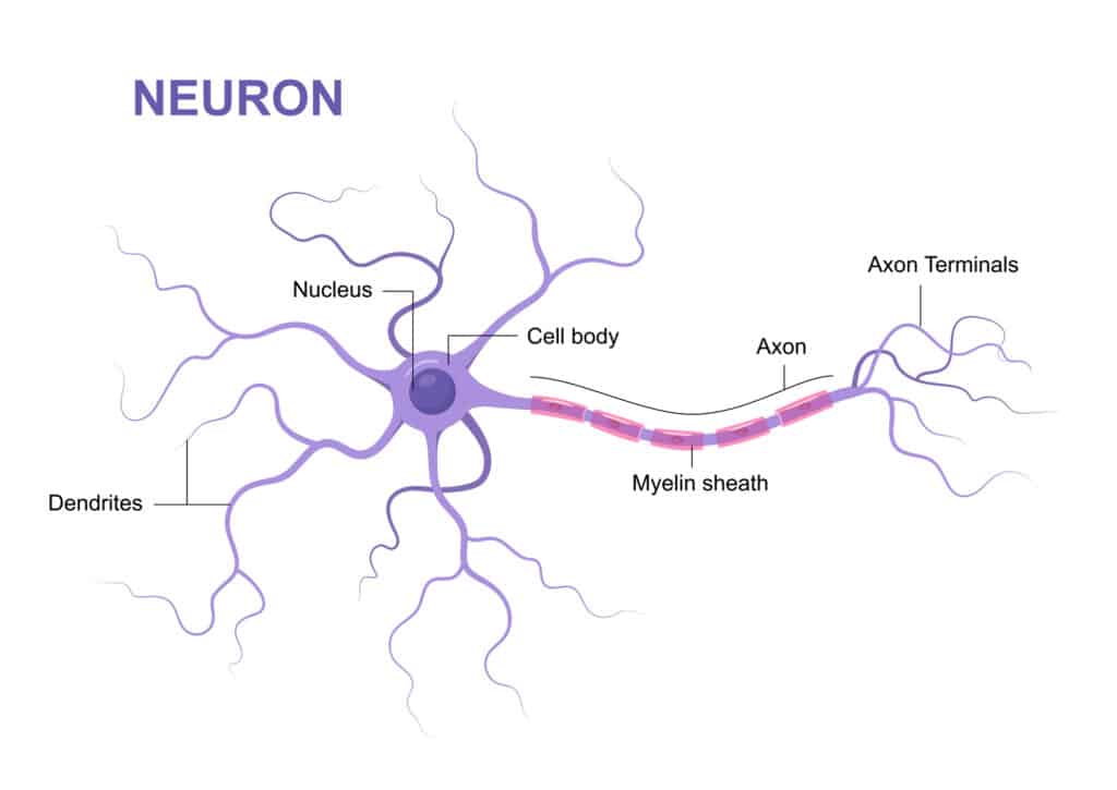
Nerve cells, also known as neurons, are basically the cells that comprise the brain and nervous system. They don’t make contact with one another directly, but connect through synapses that form between them.
Nerve cells transmit impulses along their length, and these impulses travel across synapses into other neurons. These impulses are electrical signals, known as action potentials. The action potentials need to cross multiple synaptic gaps in some cases to get to its final destination, either two or from the central nervous system.
The electrical impulse triggers the synaptic vesicles of a neuron to release neurotransmitters, which travel across the gap between the two neurons and bind to specialized receptor sites on the other neuron. This triggers the other cell to produce an action potential.
A neuron is composed of a cell body, or soma, with an axon and dendrites. The dendrites are basically little branches that can receive signals from other neurons, and the axon is a long nerve fiber that conducts electrical impulses away from the cell body. The axon is coated with a myelin sheath, an insulating layer that allows for quick transmission of the nerve impulses.
What Are Cranial Nerves?
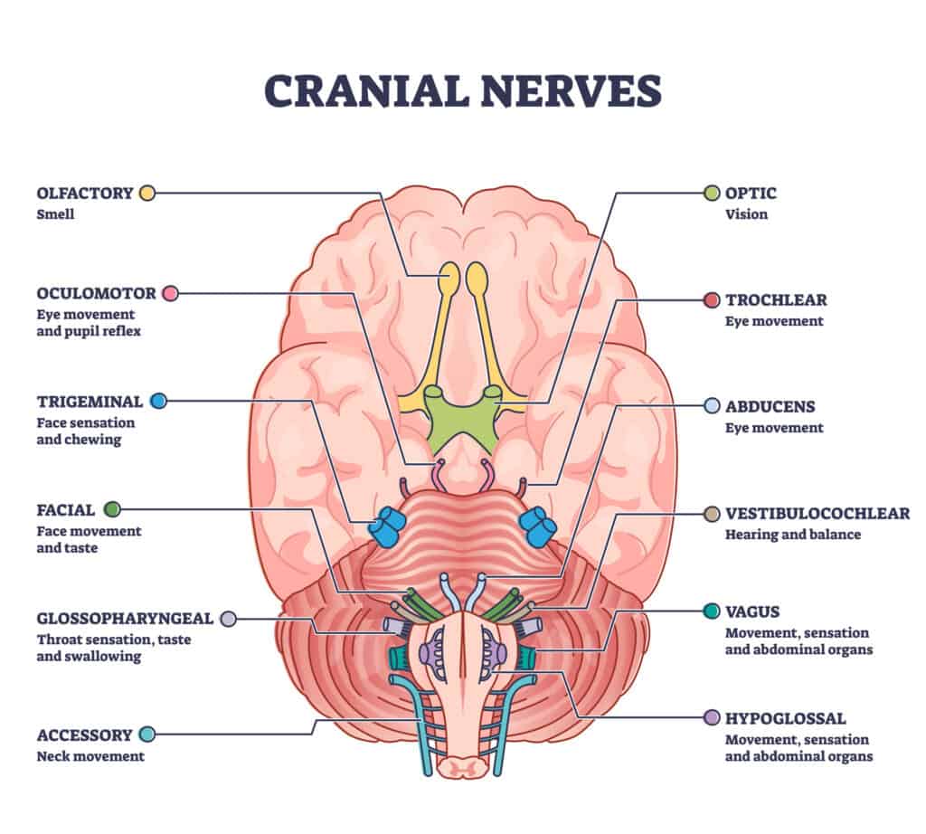
The cranial nerves are 12 pairs of nerves that send impulses to the rest of your body, allowing you to experience many sensory and motor functions.
©iStock.com/VectorMine
The cranial nerves are 12 pairs of nerves that are located in the back of your brain. They send electrical signals from your brain to other parts of your body, and vice versa. Cranial nerves make it possible for you to feel sensations, smell, hear, and taste. They also control important movements of your facial muscles.
Each of these 12 pairs of nerves splits, so that it can serve both sides of your body and brain. Each of these nerves have a specific function.
Two of these pairs originate in the cerebrum, which is the largest part of your brain. These are the optic nerves, which control your ability to see, and the olfactory nerves, which control your sense of smell. The other 10 pairs start in your brainstem, which connects your spinal cord to your brain.
The Largest Cranial Nerve
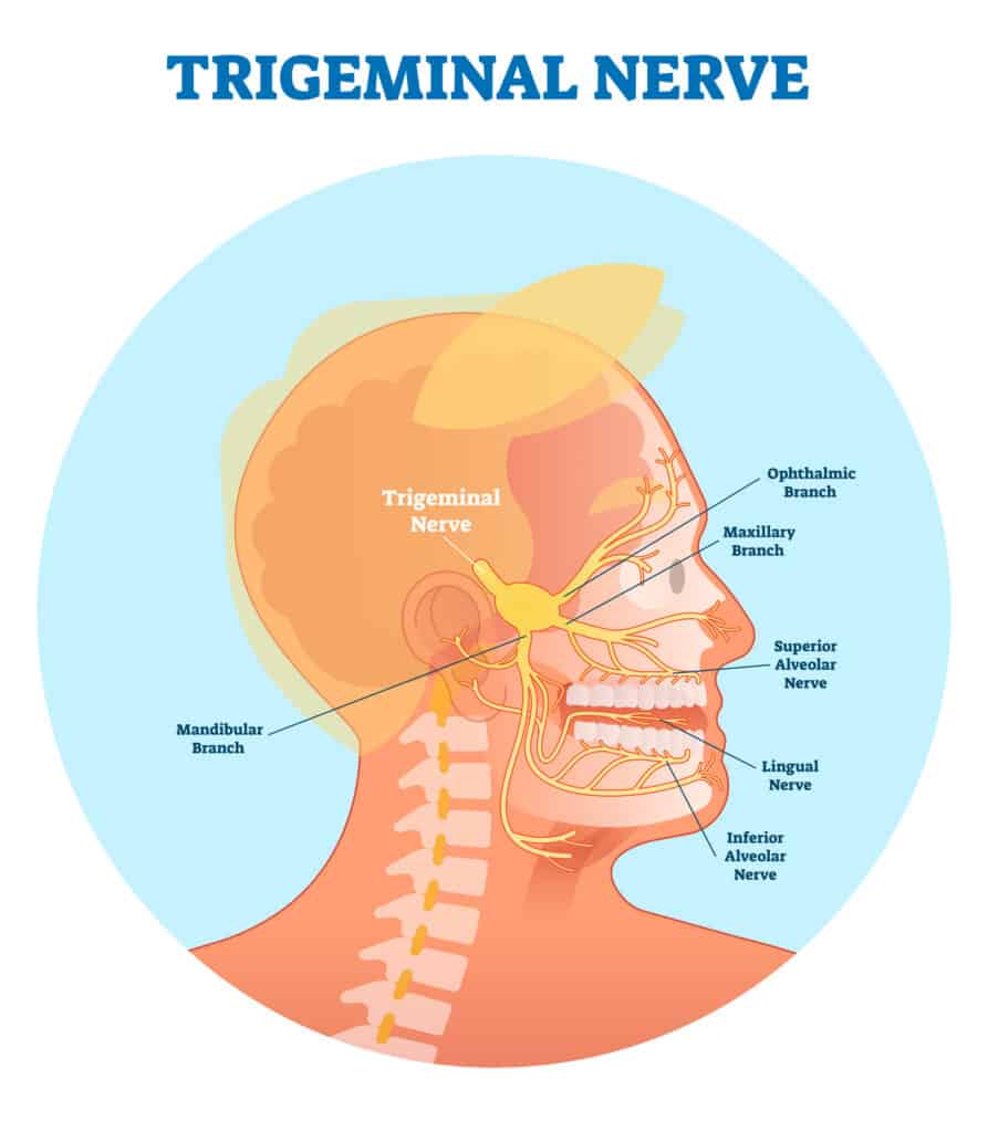
The largest cranial nerve is the trigeminal nerve, leaving the pons of your brain and forming three branches on each side that innervate different parts of your head.
©iStock.com/VectorMine
The largest cranial nerve is the trigeminal nerve. It is also the most complex of the 12 cranial nerves. It departs the brain via a large sensory root and a smaller motor root that exit the pons. The left and right trigeminal nerve branch out throughout your head.
The trigeminal nerves are the fifth of 12 cranial nerve pairs, also known as CN V. They start out with four collections of nerve cell bodies (also known as nuclei) in your brain. Three of them control your senses, and the fourth controls motor function. As they approach the pons, the three sensory nuclei combine to become one root.
Then, the sensory root splits to travel to the left and right sides of your head, becoming the trigeminal ganglia, which are located in the Meckel cave of the cranial cavity, which is close to your ear. On each side of your head, the ganglion will split into trigeminal nerve branches, which service different parts of your face. The three branches each perform an important function.
Trigeminal Nerve Branches
The ophthalmic branch (V1) transmits impulses from your scalp in the upper part of your face to your brain. This branch services your eyes, upper eyelids, and forehead.
The maxillary branch (V2) allows you to have sensations in the middle portion of your face. It travels to your gums, upper lips, lower eyelids, nose, and cheeks.
The mandibular branch (V3) helps you achieve sensation in the lower part of your face, including your gums, lower lips, and jaws. Unlike the other nerves, which only have sensory functions, these nerves also have motor functions, making it possible for you to chew and swallow.
The mandibular branch also provides the brain with sensory information from the anterior part of the tongue.
The Longest Cranial Nerve
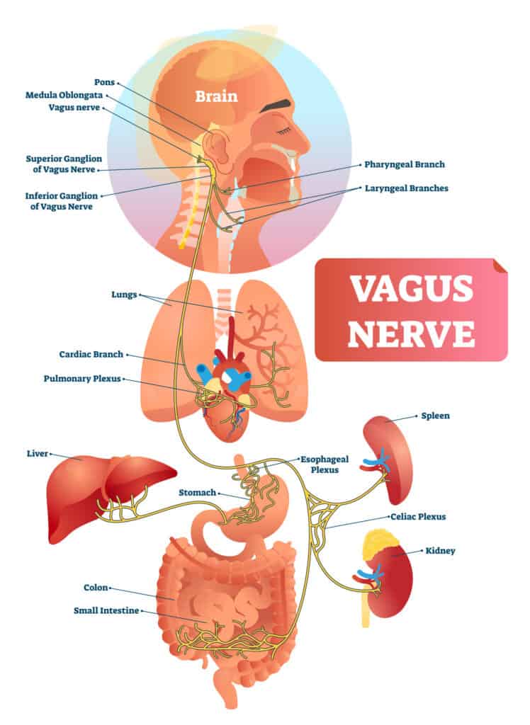
The longest cranial nerve is the vagus nerve, starting at the medulla oblongata and taking a winding course through your body before ending up at your large intestine.
©iStock.com/VectorMine
Even though the vagus nerve is not technically the largest, it is the longest of the cranial nerves. Your vagus nerves run between your brain and large intestine. The right vagus nerve goes down the right side of your body, and the left vagus nerve goes down the left side.
The vagus nerves are the 10th of 12 cranial nerve pairs, known also as CN X. They are the primary nerves of your parasympathetic nervous system, controlling involuntary body functions, including:
- Digestion
- Immune system responses
- Blood pressure and heart rate
- Urine output
- Respiration
- Sensation of the muscles and skin
- Production of saliva and mucus
- Taste
- Speech
- Mood
These nerves contain about 75% of the nerve fibers of the parasympathetic nervous system, sending information back and forth between your brain, digestive system, and heart.
These nerves actually take somewhat of a long and winding path through your body. They originate at the medulla oblongata in the lower brain stem, leaving the skull through the jugular foramen. They then connect with the neck, thorax (chest), heart, lungs, abdomen, and digestive tract before ending up at your large intestine.
The left and right vagus nerves connect together to form the vagal trunk that starts to go down your body. They connect at the point where your esophagus joins your abdominal cavity, which is known as the esophageal hiatus. The vagal trunk includes gastric nerves that travel to your abdomen.
There are three major vagal nerve branches. The vagus nerve branch interacts with nerves that go to your lungs, heart, and esophagus. The superior ganglion branch interacts with nerves that go to your ear and spine. The inferior ganglion branch interacts with nerves and muscles that go to your larynx (voice box) and pharynx (throat).
The photo featured at the top of this post is © iStock.com/mstroz


How to Add Us to Google News


Thank you for reading! Have some feedback for us? Contact the AZ Animals editorial team.