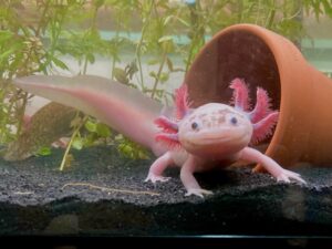
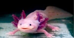
Axolotls (Ambystoma mexicanum), also known as the Mexican salamander or the Mexican walking fish, are aquatic amphibians that belong to the family Ambystomatidae. Axolotls are famous for their ability to regenerate lost body parts, including their vestigial teeth. Vestigial teeth are teeth that have lost their original function through evolution and are no longer useful to the organism.
The vestigial teeth of axolotls are thought to be a remnant of their evolutionary history. Axolotls are believed to have descended from salamanders that had functional teeth, which were used for grasping and tearing food. However, as axolotls evolved to feed on smaller prey, their teeth slowly became smaller and less useable. Eventually, they became vestigial structures.
Despite their small size and lack of function, axolotl teeth are still an important feature of the anatomy of this animal. Scientists use the presence and position of teeth in axolotls to identify different species and subspecies. In addition, scientists use them to study the evolutionary history of these fascinating animals. Keep reading to learn more about these captivatingly adorable amphibians.
Axolotl Larval Teeth
The axolotl larvae have teeth that are used for feeding on small organisms in their aquatic environment. Axolotl larvae usually have multiple rows of small, cone-shaped teeth on each side of their upper and lower jaws, which are shed and replaced often as the larvae grow. These teeth are relatively simple in structure and function and are not clearly differentiated or delineated.
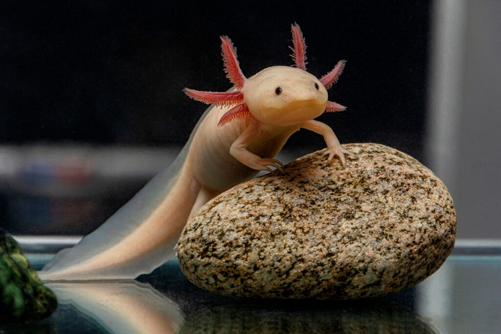
Despite their small size and lack of function, axolotl teeth are still an important feature of the animal’s anatomy.
©Iva Dimova/Shutterstock.com
What Kind of Teeth Do Adult Axolotls Have?
Adult Axolotls once had fully formed, functional teeth in their evolutionary history. However, over time, these teeth became smaller and less useful as the axolotl adapted to its environment and changes in its diet.
Axolotls evolved from a lineage of aquatic ancestors that had teeth that were well-suited for catching and eating prey in water. As the axolotl evolved, it became adapted to a different diet. The new diet consisted of consisting primarily of soft-bodied prey. Therefore, they no longer require the use of sharp, pointed teeth for biting or chewing. However, axolotl mouths continue to harbor the remnants of their former teeth. They have between 30-40 teeth in each jaw, which are small and inconspicuous. The teeth are not used for chewing, but rather for holding small prey items as the axolotl sucks them into its mouth.
The principal areas of interest in the axolotl mouth are:
- outer dental arcade, which contains the:
- premaxillary tooth field
- maxillary tooth field
- dentary tooth field
- inner dental arcade, which contains the:
- vomerine tooth field
- palatine tooth field
- coronoid (or splenial) tooth field
Axolotl Teeth: Outer Dental Arcade
The outer dental arcade is the outermost row of teeth. It contains the premaxillary, maxillary, and also dentary tooth fields. The teeth in the outer dental arcade are smaller and less prominent than those in the inner dental arcade. They are arranged in a single row. During feeding the teeth in the outer dental arcade are particularly important for snagging and immobilizing small, fast-moving prey, such as insects and small fish.
Premaxillary Teeth
Premaxillary teeth are the smaller teeth located at the very front of the upper jaw. These teeth are typically smaller and more pointed than the maxillary teeth. They are arranged in a single row. The premaxillary teeth are used for grasping and holding prey, particularly small invertebrates.
Maxillary Teeth
Maxillary teeth are the larger and more numerous teeth located on the maxilla bone, which forms the bulk of the upper jaw. These teeth are larger and are arranged in multiple rows. These teeth can be regenerated and also replaced throughout the axolotl’s life.
Dentary Tooth Field
The dentary tooth field is located in the lower jaw and contains small, pointed teeth that are used for grasping and holding onto prey. The teeth in the dentary tooth field are arranged in several rows along the outer edge of the lower jaw and are often smaller and less prominent than the maxillary teeth in the upper jaw. These teeth work in conjunction with the maxillary teeth in the upper jaw to immobilize and hold onto prey while the axolotl manipulates the prey item with its tongue and throat muscles to move it toward the back of the mouth for swallowing. The dentary teeth, like other teeth in the axolotl’s mouth, are continuously replaced throughout the animal’s life. This ensures that it always has a fresh supply of functional teeth for prey capture.
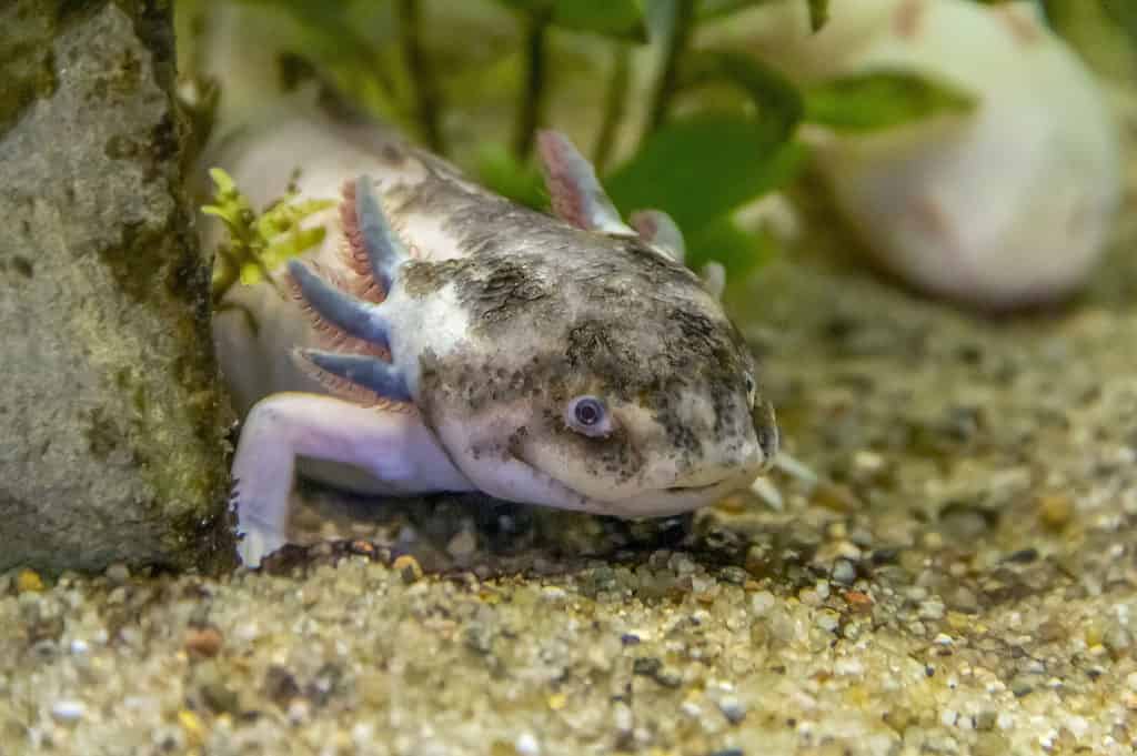
Axolotls have between 30-40 teeth in each jaw, which are small and hidden.
©iStock.com/prill
Axolotl Teeth: Inner Dental Arcade
The inner dental arcade is the row of teeth located in the upper jaw and the palate. The inner dental arcade contains the vomerine, palatine, and also coronoid tooth fields. The teeth in the inner dental arcade are larger and more numerous than those in the outer dental arcade. The teeth in the inner dental arcade are arranged in multiple rows along the edge of each maxilla bone; They are conical with a dull point on the tip.
Vomerine Tooth Field
The vomerine tooth field is located on the vomer bone, which forms part of the nasal septum in the roof of the mouth. The teeth in this field are relatively small and are used for grabbing and holding prey, particularly small invertebrates. The vomerine teeth are arranged in a V-shaped pattern, with the apex of the V pointing toward the back of the mouth. In axolotls, the vomerine teeth are present at birth and do not regenerate throughout the life of the animal.
Palatine Tooth Field
The palatine tooth field is located on the palatine bones, which form the roof of the mouth. The teeth in this field are larger and more prominent than the vomerine teeth. The palatine teeth are arranged in several rows, with the largest teeth located in the center and the smaller teeth arranged in a semicircle around them. The palatine teeth are continuously replaced throughout the axolotl’s life.
Coronoid Tooth Field
The coronoid (or splenial) tooth field is located on the lower jaw bone, known as the coronoid or splenial bone. The teeth in this field are also relatively small and are used for grabbing and holding prey. The coronoid teeth are arranged along the edge of the coronoid bone. The coronoid teeth do not regenerate throughout the life of this animal.
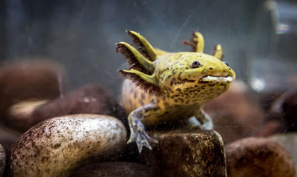
By copying the natural processes of tooth regeneration and the structure of axolotl teeth, researchers hope to develop new solutions for human dental problems and other medical challenges.
©iStock.com/Gerardo Martinez Cons
Biomimicry and Axolotl Teeth
Once research concluded that they regenerate their teeth throughout their lives, studying the mechanisms behind tooth regeneration in axolotls become a field in biomimetics. Biomimicry refers to the practice of using natural biological systems and processes as inspiration for developing new technologies, materials, and designs. In this case, scientists are studying the unique properties of axolotl teeth and the mechanisms behind tooth regeneration in order to develop new dental treatments and materials for medical applications. By copying the natural ways of tooth regeneration and the structure of axolotl teeth, researchers hope to develop innovative solutions for human dental problems and other medical challenges.
Recently, researchers have identified several genes and signaling pathways that are involved in tooth regeneration in axolotls. These discoveries have opened up new avenues of research into regenerative medicine and dental therapies. Scientists are now exploring ways to apply the knowledge gained from axolotl tooth regeneration to develop new treatments for tooth decay, gum disease, and other dental problems in humans.
The photo featured at the top of this post is © axolotlowner/Shutterstock.com
FAQs (Frequently Asked Questions)
What is an axolotl?
Axolotls, also known as Mexican salamanders, or Mexican walking fish, are aquatic amphibians that belong to the family Ambystomatidae. While axolotls are known for their ability to regenerate lost body parts, they also have vestigial teeth, which are teeth that have lost their original function through evolution and are no longer useful to the organism.
What are vestigial teeth?
Vestigial teeth are small, poorly developed, and not functional in the sense that they are not used for grinding or tearing prey. The loss of functional teeth in axolotls is thought to be an evolutionry adaptation to their unique environment and feeding habits. Axolotls live in shallow, freshwater environments where there is often an abundance of small, soft-bodied prey, such as insects and crustaceans. These types of prey do not require a lot of biting or chewing to be digested, so the loss of teeth has not been a major disadvantage for the axolotl.
Where do axolotls live?
Axolotls are native to the freshwater canals and lakes surrounding Mexico City, Mexico. Specifically, they are found in the Xochimilco and Chalco regions, which are located in the Southern part of the city.
In the wild, axolotls are primarily found in the shallow waters of these canals and lakes, where they feed on a variety of small prey items such as insects, small fish, and crustaceans. They are well-adapted to life in freshwater environments, with specialized gills that allow them to extract oxygen from the water and a streamlined body shape that helps them move efficiently through the water.
Do axolotls make good pets?
Absolutely! These enchanting amphibians make splendid pets! Axolotls are commonly kept in captivity as pets and are popular among hobbyists and researchers due to their unique physiology and regenerative abilities. Their lack of functional teeth makes them relatively safe aquatic pets for young children.
How many different species of axolotl are there?
There is currently only one recognized species of axolotl, which is known scientifically as Ambystoma mexicanum. This species is the only member of its genus and is one of several species of salamander found in Mexico and parts of the Southwestern United States.
While there is only one recognized species of axolotl, there is some variation in appearance and coloration between individuals from different regions of their native range. However, these differences are generally considered to be within the normal range of variation for the species and do not represent distinct subspecies or varieties.
Thank you for reading! Have some feedback for us? Contact the AZ Animals editorial team.


