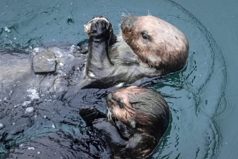

Chordates are animals that belong to the phylum Chordata. These animals are unified by the presence in their bodies of a flexible rod that supports their backsides at some point during their development. While most chordates are vertebrates and have backbones, some are invertebrates.
Characteristics of Chordates
Chordates take an incredible variety of forms. For example, we humans share the phylum Chordata with such drastically different animals as sea squirts and lancelets! While you might not guess it by looking at them, they share several traits with us that don’t appear in non-chordate animals. They are as follows:
- A notochord — from the Greek noto-, meaning back, and -chord, meaning string. This flexible rod is the defining trait for which the phylum Chordata is named.
- In vertebrate chordates like fish, birds, mammals, reptiles, and amphibians, the notochord is present only during the embryo stage. Here, it supports the embryo as it develops and begins the early formation of the central nervous system. The vertebral column, or spine, replaces it as a support structure for the animal’s skeletal system at later developmental stages.
- Chordata contains two invertebrate groups: the tunicates of subphylum Urochordata and the lancelets of Cephalochordata. These animals never develop a spinal column and instead retain their notochord throughout their lives.
- A dorsal hollow nerve tube — This begins as the primitive central nervous system that develops in the embryo above the notochord. The tube wall thickens as it develops, leaving a narrow but clearly defined cavity that runs its length. Hollow nerve tubes are present in all chordates, both vertebrate and invertebrate, and attach to nerve tissues throughout the body.
- Pharyngeal slits — All chordate embryos have a set of slits behind the pharynx, which is the part of the digestive tract that connects the mouth to the esophagus. Not all adult chordates retain these slits.
- In many aquatic species, these ultimately develop into the gill slits or parts of the jaw. In suspension-feeding animals, the pharyngeal slits are lined with cilia. These pull water into the mouth and through the slits, capturing food particles as they do so.
- In terrestrial animals, the embryonic pharyngeal slits develop into completely different structures, such as components of the jaw and inner ear. In many terrestrial animals, including humans, the pharyngeal slits also develop into the tonsils.
- A post-anal tail — All embryonic chordates have a part of the notochord that extends past the anus. While some species retain this feature into adulthood, many, like humans and frogs, lose it entirely.
Examples of Chordates
While many of the characteristics of chordates are present only in the embryonic stage, they can manifest as a diverse array of structures by adulthood. This incredible diversity in development can be highlighted by comparing animal species to the lancelets, who retain all of the basic characteristics of chordates into adulthood.
Let’s have a look at the anatomy of a lancelet:
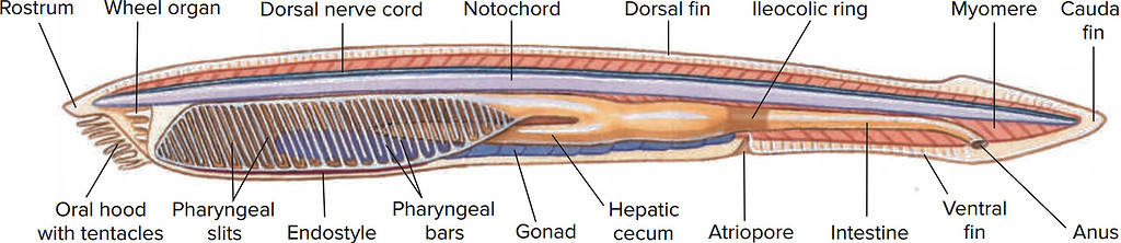
Adult lancelets retain all the qualities of chordates into adulthood. Their anatomy and genome can give biologists strong clues about the origins and evolution of vertebrates.
Note the long notochord that runs along the lancelet’s entire backside and extends past the anus. Just above it is the nerve cord that sends signals throughout its body. Behind the oral hood, which acts as a coarse pre-filter, the pharyngeal slits allow the lancelet to filter out smaller, edible food particles from the surrounding water.
Now that we have a good example of what these structures look like, let’s take a look at some of the ways they develop differently in other animals.
Notochord
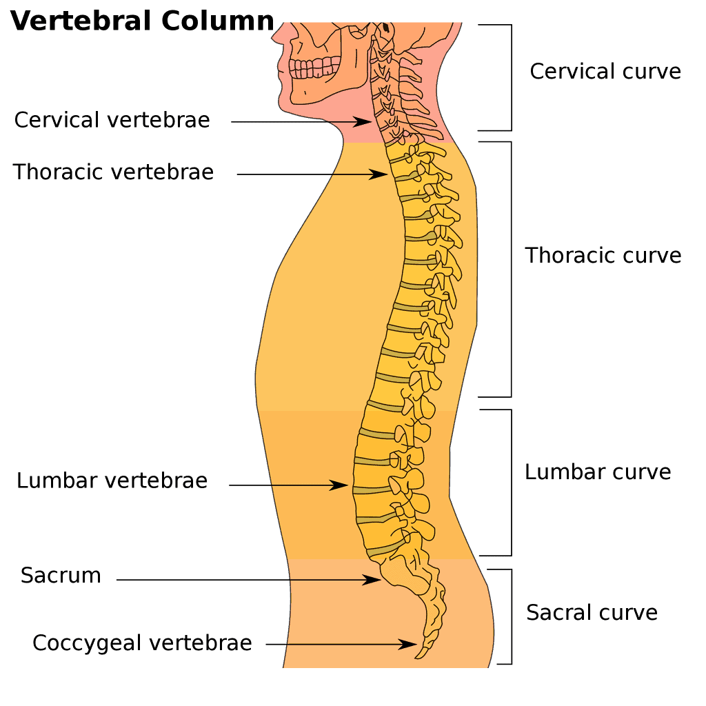
Rather than develop a more complex support structure, lancelets retain their notochords throughout their lives. In other animals, however, the notochord gives rise to the vertebral column later on in development. Vertebral columns vary in overall length and structure, but all are comprised of many segments — vertebrae — that support the body, protect the spinal cord, and allow complex movements. Compare the simple notochord of the lancelet to the spinal column of the goat below. The goat’s vertebral column, like our own, is divided into several regions of vertebrae based on location, form, and function.
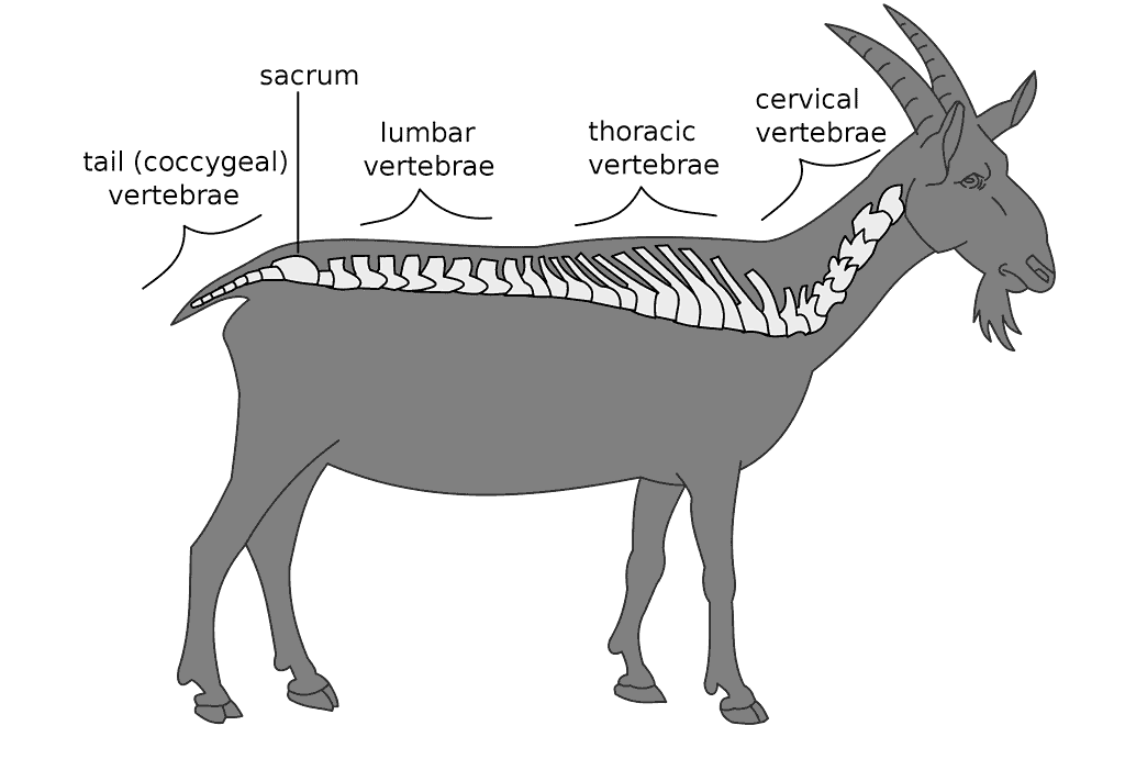
The vertebral column of the goat, like our own, develops from its embryonic notochord. Inside it, the hollow nerve tube becomes the spinal cord, forming part of the central nervous system.
Pharyngeal Slits
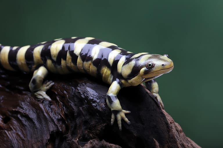
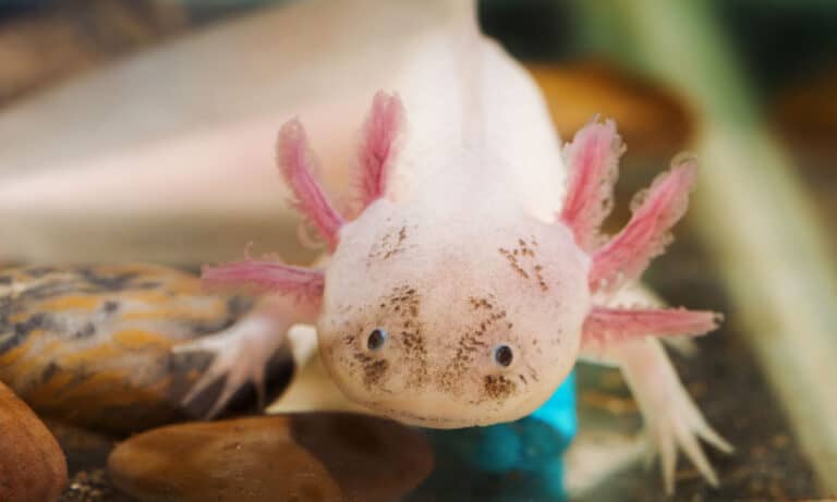
While a lancelet’s pharyngeal slits serve to isolate food from passing water, they develop further and perform different functions in other animals. In fish, for example, the pharyngeal slits become the gills and gill slits, which scour oxygen from the surrounding water. They become gill slits in amphibians as well, though many species lose them by adulthood.
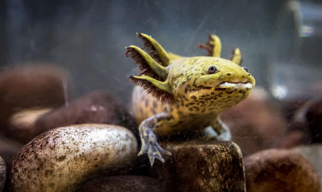
The axolotl’s gill slits rest behind its feathery external gills, helping pass water over their undersides.
©iStock.com/Gerardo Martinez Cons
One species of salamander, the axolotl (Ambystoma mexicanum), never undergoes complete metamorphosis and retains its gill slits throughout its life. Its gills themselves, however, are external. Inside the gill slits are rakers that allow oxygen-rich water to pass over the gills, but restrict larger particles that may damage the sensitive gill tissue. The rakers also prevent food from escaping the salamander’s mouth. In this case, the gill slits carry out similar functions to those of the pharyngeal slits with the addition of respiration.
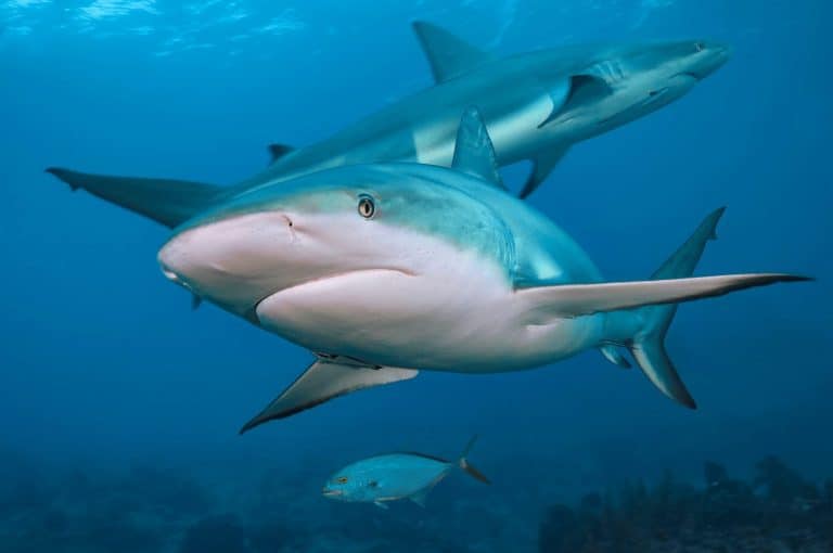
In fish, on the other hand, the pharyngeal slits develop into sets of internal gills. These consist of pharyngeal arches that bear very fine, filaments of gill tissue. The dense network of blood vessels within the gill tissue allows the fish to exchange gases like oxygen and carbon dioxide with the water around it. Unlike the axolotl which has external gills, fish have various types of coverings that protect their gills from the outside world. Bony fish, like tuna, have a single gill opening on each side of their bodies. These are each guarded by a hard, bony plate called the operculum. Sharks and rays, on the other hand, have several gill openings on each side that are covered by thickened skin.
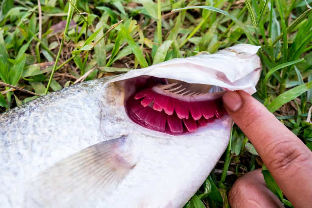
Bony fish like this snapper have internal gills. The red, filamentous gill tissue allows the fish to absorb oxygen from the surrounding water while the bony gill rakers prevent large objects from passing through. The hard operculum protects the entire assembly.
©koliw/Shutterstock.com
Post-Anal Tail
At some point in their development, all chordates have a portion of their support column that extends past the anus. You can see the post-anal tail in the diagram of the lancelet above as the notochord stretches out to form the caudal fin. It is also visible in the drawing of the goat, whose coccygeal spine forms its bony tail. While these two chordates retain their post-anal tails throughout their lives, there are some who lose it by adulthood.
Humans are a great and familiar example. Though we possess a protruding tail during our early development, it disappears by the time our embryos are eight weeks old. All we are left with by the time we are born is a small nub. This nub — the coccyx, or tailbone — serves as an attachment point for various muscles in the lower body. Otherwise, it is a reminder of our evolutionary past when we would have used our tails for signaling or balance.
The very last vertebrae in the human spinal column, the coccygeal vertebrae, are all that remain of our post-anal tails.
Humans aren’t the only chordates to grow out of their post-anal tails. Frogs do this as well. When they first emerge from their eggs, tadpoles are very simple. They have a mouth to eat, gills to breathe, and a tail to move around. As they grow, they develop teeth, two pairs of legs, and a long spinal column. Eventually, they lose their gills and develop lungs instead. During the froglet stage, they look just like adult frogs with long tails, which they gradually reabsorb. By about 14 weeks of age, the tail disappears entirely, leaving behind it only a nub reminiscent of our own tailbones.
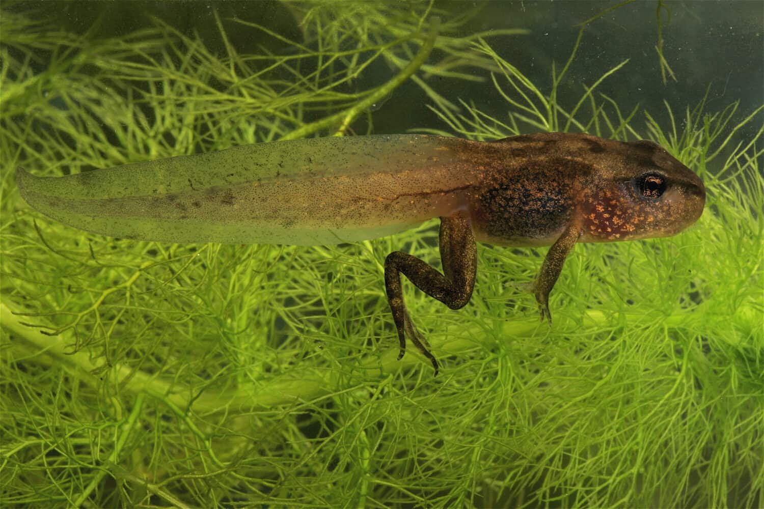
Soon, this little froglet will completely reabsorb its post-anal tail and emerge from the water as an adult frog.
©W. de Vries/Shutterstock.com