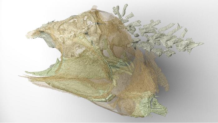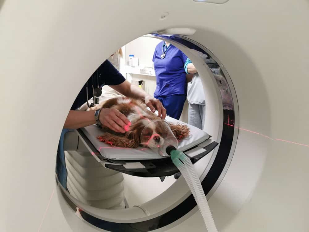

Several imaging procedures have been developed to help diagnose diseases in humans. Examples include X-ray imaging, ultrasonography, and computed tomography, also known as CT scans. CT scans have been adapted for use in animals. They can provide a lot of information without the need for invasive procedures or altering the disease.
However, they do require highly specialized equipment, skilled operators, and animals that need to keep very still! In this fascinating Instagram post, you can see a staggering variety of animals of all sizes being prepared for and receiving CT scans. Kittens, hamsters, horses, dogs, lions, birds, and even fish are undergoing the procedure. Here we explain exactly what a CT scan is, when it is used, and how animals are prepared for the procedure.
How Do CT Scans Work?
CT scans are essentially a series of computer-controlled X-rays. The animal (or person) is placed on a motorized bed that moves into the scanner. One X-ray is taken, and then the bed is moved very slightly forward. A computer uses multiple X-rays to produce a 3D image. The scan creates cross-sectional images of structures and organs within the body, providing a more detailed view. The images are displayed on a monitor. If needed, a dye (called contrast), which is visible on X-rays, can be injected intravenously to detect structural changes deep within the body. The crucial thing is that for a CT scan to be conducted successfully, the patient must be still throughout.
The Advantages of CT Scans
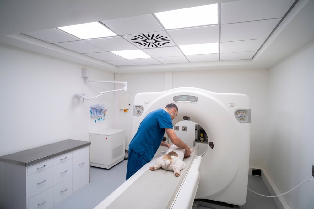
CT scans are very quick.
©Carrastock/Shutterstock.com
Modern multi-slice CT scanners can acquire thousands of images in seconds. The scanner rotates continuously around the patient, and this is coordinated with the movement of their bed. For small animals, it takes less than 10 seconds to get a complete scan of their abdomen or chest. Some scanners can even capture scans of their beating heart during all stages of the cardiac cycle.
Speed is the greatest advantage of CT scans. An entire 3-D picture of an organ can be obtained in the same time that it takes to capture a single X-ray! Amazingly, a 64-slice scanner can generate over 5,000 images in just 10 minutes, which the computer then compiles into a single comprehensive scan.
What Are CT Scans Used For?
Vets use CT scans to look at deep tissue and organs that are not visible. They are used to obtain images of the brain, spinal cord, and lungs, for example. Animals may receive CT scans if they are lame, as the scans can detect anomalies in the elbows, shoulders, and limbs. They are also useful for nasal investigations, ear problems, brain disease, and suspected cancers.
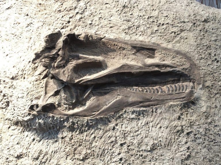
CT scans of the skull have been especially useful because they reveal the complex anatomy of the skull, which has lots of overlying structures. This makes a diagnosis much more accurate and specific. It can be a useful diagnostic method for spinal cord disease in small animals and is also very useful in cancer diagnoses. These scans can detect small metastatic (secondary) tumors. They have also been used to guide biopsy and aspirate sampling procedures and to accurately target radiation therapy.
Even structural changes deep within the body can be detected. Tumors, abscesses, vascular abnormalities, occult fractures, and hematomas have all been detected using these scans. Not everyone can interpret a CT scan, however! You need an expert level of knowledge about the anatomy of the scanned animal. Every animal species is different! Scan reviewers must be able to identify all the structures in any plane through the body so that they can spot changes in anatomy or physiology. Specific training is therefore required.
Preparing Animals for CT Scans
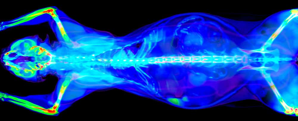
Cancer can be diagnosed with CT scans.
©Benny Marty/Shutterstock.com
CT scans may be quick, but they are not fast enough to capture images of a wriggling dog or cat or a swaying horse! So, veterinary patients must be anesthetized and immobilized for the procedure. That said, the period of anesthesia is short and is not too risky for most animals.
Researchers have attempted to capture CT scans of fully awake or minimally sedated animals, but the lack of controlled positioning resulted in a lot of movement! Consequently, the images were difficult to interpret, so sedation is always preferred. That said, for a few animals, it is safer to confine them physically than to anesthetize them. Take the hamsters, for example. Anesthesia comes with a higher risk for these little pets. The best option, therefore, is to create a hamster burrito by binding them quite tightly. This ensures that they cannot move, but it is far less risky. The same is done for young kittens. An added advantage is that two can be scanned at once, and they look especially cute during the process!