
Quick Take
- For decades, early fossil fish appeared to have bizarrely tiny brains inside enormous skulls. The real explanation for why involves a subtle scanning mistake that changes how paleontologists read fossils. Why tiny brains confused scientists →
- The only reason a 300-million-year-old brain survived at all comes down to a chemical quirk nicknamed 'fool's gold,' and by all accounts it shouldn't have worked. How fool's gold preserved the brain →
- One corner of this ancient fish's brain shares a distinctive pattern with fish still alive today, a finding that may finally settle a long-running argument about which modern lineage they belong to. Cerebellum clues to fish lineage →
- Researchers couldn't touch this fossil without destroying it, so they found a way to read it anyway. Their approach unlocked details that traditional lab methods had missed for years. How CT scanning unlocked the fossil →
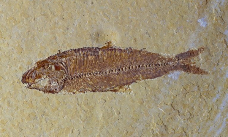
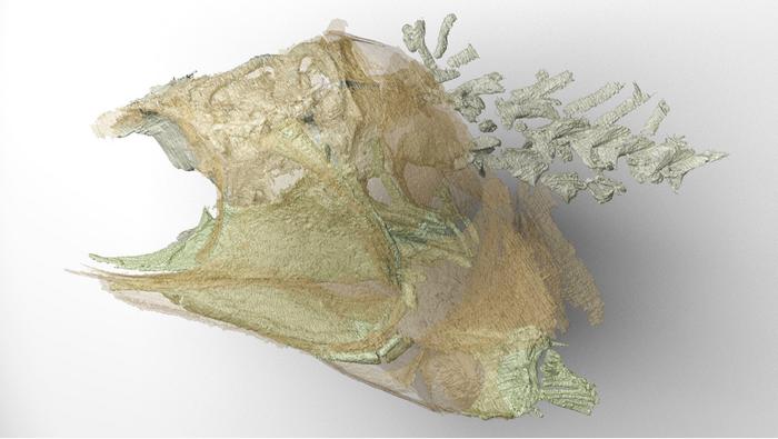
More than 300 million years ago, a small ray-finned fish died in what is now northwest England and settled into the sediments of a prehistoric swamp. Over immense spans of time, the remains of that fish — now known as Trawdenia planti — became fossilized in a layer of soapstone between coal seams in the Burnley coalfields.
What makes this fossil extraordinary is not just its skeleton, but the preservation of delicate neural tissue from the brain. In a new study published in the Proceedings of the National Academy of Sciences (DOI: 10.1073/pnas.2610438123), researchers at the University of Chicago used modern imaging techniques to examine the fossil and uncover clues about the early evolution of ray-finned fishes, the group that includes the overwhelming majority of fish alive today.

Dr. Coates is the senior author of the study and Professor and Chair of Organismal Biology and Anatomy at the University of Chicago.
Why Preserved Brain Tissue Is So Unusual
Neural tissue is rarely preserved in fossils because it is extremely delicate and decays rapidly after death. Soft tissue preservation is uncommon in general, but brain tissue is especially unlikely to survive long enough to leave a meaningful fossil record.
Researchers believe Trawdenia was preserved through a highly specific set of chemical conditions. “Neural membrane tissues may have undergone cross-linking between proteins and metal ions — especially iron — leading to pyritization, sometimes called “fool’s gold.” Similar preservation has been observed before in fossil invertebrates, but the process remains exceptional.”
Dr. Michael Coates
What CT Scanning Made Possible
The fossil could not simply be prepared out of the surrounding rock by hand. The skull and soft tissues were partly encased within a nodule of clay-rich ironstone, and the structures were far too delicate to be removed mechanically without being destroyed.
CT scanning was essential because it allowed researchers to image the fossil inside the rock. This enabled them to study the skull and internal soft-tissue structures without damaging the specimen, revealing anatomical details that would have been inaccessible through traditional preparation methods.
Why the Brain’s “Snug Fit” Matters
One of the most important findings in the study is that the preserved brain appears to fit closely within the braincase.
This is important because earlier CT-based studies of fossil brains in some early fishes identified structures that appeared unusually small compared to the large cavities of the skull. This raised an obvious question: why would a fish have such a large braincase surrounding such a tiny brain?
This study offers a possible answer. Coates explains that earlier scans may have detected only the inner chambers of the brain — the ventricles, which held cerebrospinal fluid during life — rather than the full bulk of the brain itself. The researchers initially detected a similar signal in Trawdenia, but further analysis revealed evidence for the broader body of the brain as well.
If this interpretation is correct, it changes how paleontologists interpret fossil braincases. The natural mold inside the skull may preserve the general shape of the brain and its major subdivisions to a reasonable extent, even when soft tissue is not fully preserved.
A Clue to the Origins of Modern Fish Groups
The fossil also offers insight into where Trawdenia may fit among early ray-finned fishes. In this study, the researchers found that the way major brain divisions are packed together can be characteristic of different natural groups of fishes.
One feature that links Trawdenia to modern sturgeons and paddlefish is the cerebellum, which in these fishes wraps around the sides of the midbrain in a distinctive way. The new fossil appears to share that pattern, suggesting a possible deep relationship to that lineage.
Coates notes that further research is needed on this question, but the results highlight the value of brain anatomy as a source of evolutionary evidence in groups where relationships have long remained uncertain.
Why Early Ray-Finned Fish Evolution Has Been Hard to Untangle
Ray-finned fishes have a vast fossil record, but their early evolution has been difficult to resolve. Coates attributes this largely to a simple problem: relatively few researchers, an enormous number of fossils, and a longstanding lack of detailed anatomical data from the earliest members of the group.
That is beginning to change. With newer imaging tools, researchers can now recover types of anatomical evidence previously hidden inside rock or overlooked in older scans. Fossils like Trawdenia planti demonstrate how much information may still be hidden inside long-known specimens — and how modern technology can help reveal it.