
Quick Take
- Scientists used a powerful synchrotron X-ray system, robotics, and AI to scan 2,000 ant specimens in just one week, creating 3D models representing 800 species.
- The models reveal astonishing internal detail, including muscles, nerves, digestive systems, and stingers, allowing scientists to study ant anatomy without destroying specimens.
- Researchers say the open-access Antscan digital library could transform biodiversity science, education, and even animation by making detailed models of organisms widely available.
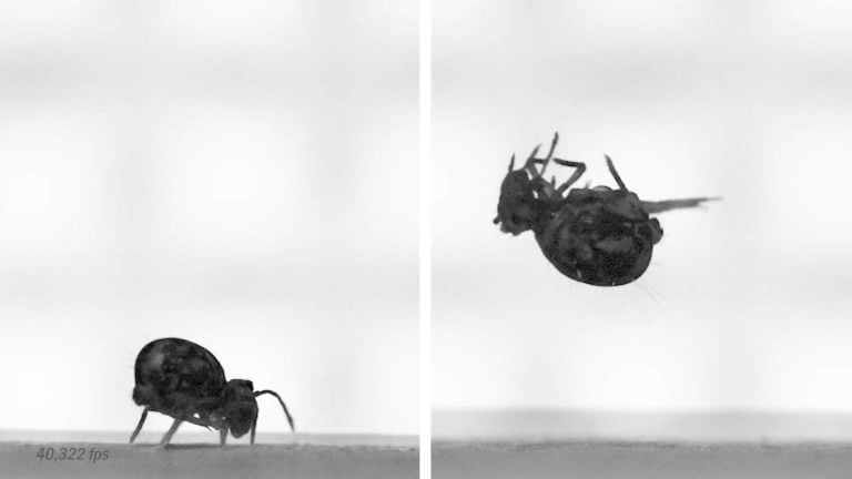
Scientists estimate there may be more than 20 quadrillion ants on Earth. Yet despite their abundance, studying the fine details of ant anatomy has always been slow work. Each specimen typically has to be examined under microscopes or scanned individually with specialized imaging equipment. That process can take hours for a single insect.
But researchers have just sped things up dramatically. An international team of scientists has created a massive new digital library of ants using high-powered X-ray scanning, robotics, and artificial intelligence. Their project, called Antscan, produced interactive 3D models representing 800 ant species by scanning 2,000 specimens in just one week.
The models are detailed enough to reveal internal features such as muscles, nerves, digestive systems, and stingers. Even more importantly, the entire dataset is freely available online, allowing scientists, educators, and artists to explore the anatomy of ants in ways that simply weren’t possible before.
Beyond ants, researchers say this approach could transform how scientists study biodiversity across the planet.
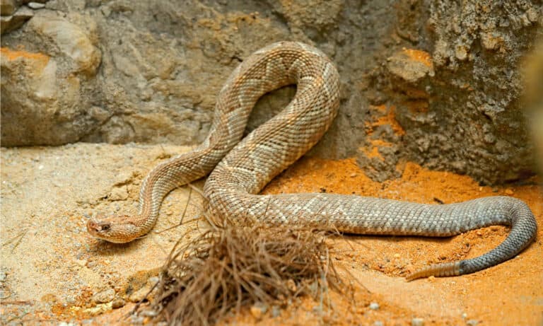
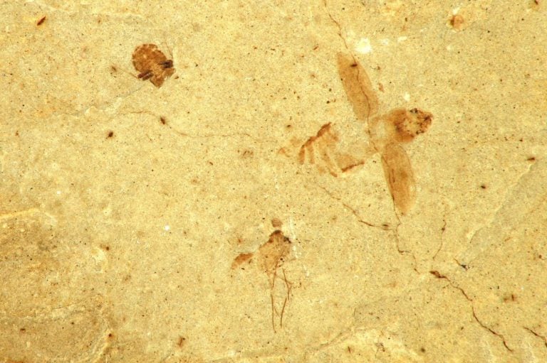
A traditional micro-CT scan of a single insect can take around 10 hours, which is why researchers needed a faster way to digitize thousands of specimens.
©Eric Isselee/Shutterstock.com
Scanning Tiny Creatures
For more than a decade, evolutionary biologist Evan Economo and his colleagues have relied on micro-CT scanning to study insect anatomy. Micro-CT machines work somewhat like the medical CT scanners used in hospitals. Instead of creating images of the human body, they produce extremely detailed X-ray images of tiny objects such as insects.
These scanners capture hundreds or thousands of cross-sectional images. Computer software then stacks those slices together to build a three-dimensional model. For entomologists studying insect form and structure, the technique has been revolutionary.
But there’s a major drawback; It’s slow.
Scanning a single insect specimen using a typical laboratory micro-CT system can take about ten hours. That means building a large dataset could take years. According to researchers involved in Antscan, scanning thousands of specimens using traditional equipment might require roughly six years of continuous operation.
Enter the synchrotron particle accelerator…
Harnessing the Power of a Synchrotron
Synchrotrons are enormous research facilities designed to accelerate particles close to the speed of light. As those particles move through curved paths, they emit extremely bright beams of X-rays, which scientists often to use to study the atomic structure of materials, proteins, fossils, and other objects.
For the Antscan project, researchers used the synchrotron at the Karlsruhe Institute of Technology in Germany. Compared with standard micro-CT systems, synchrotron X-rays are vastly brighter and more focused, allowing specimens to be scanned far more quickly while still capturing extremely fine detail.
The team also employed automated robotics. A robotic sample changer continuously rotated and swapped out ant specimens during scanning. Each specimen could be replaced in about 30 seconds, allowing hundreds or even thousands of scans to happen in rapid succession.
Compare that to the 10 hours of the previous method! This automated pipeline allowed the researchers to capture images of roughly 2,000 ant specimens in just a single week—a dramatic leap compared with traditional lab-based CT scanning.
The resulting images were incredibly detailed—sharp enough to show structures only a few millionths of a yard across. That level of resolution reveals features that are normally hidden even under high-powered microscopes.
From Raw Scans to Living Models
Creating high-resolution scans is only the first step in building a digital organism library. Raw CT scans often show specimens in awkward or distorted positions. Ants preserved in alcohol for museum collections frequently curl up or bend their legs in unnatural ways.
To transform those scans into realistic digital insects, the team turned to artificial intelligence.
Computer science students at the University of Maryland began developing AI tools capable of performing “pose estimation.” This process uses machine learning algorithms to identify anatomical landmarks and reposition body parts into more natural poses.
Instead of manually adjusting every leg, antenna, and body segment, the AI system can automatically reconstruct an ant in a lifelike posture. The result is an interactive 3D model that resembles how the insect would appear when walking through its natural environment.
These models can be rotated, zoomed, and explored from any angle. Researchers can even digitally slice through the ant to view internal structures layer by layer.
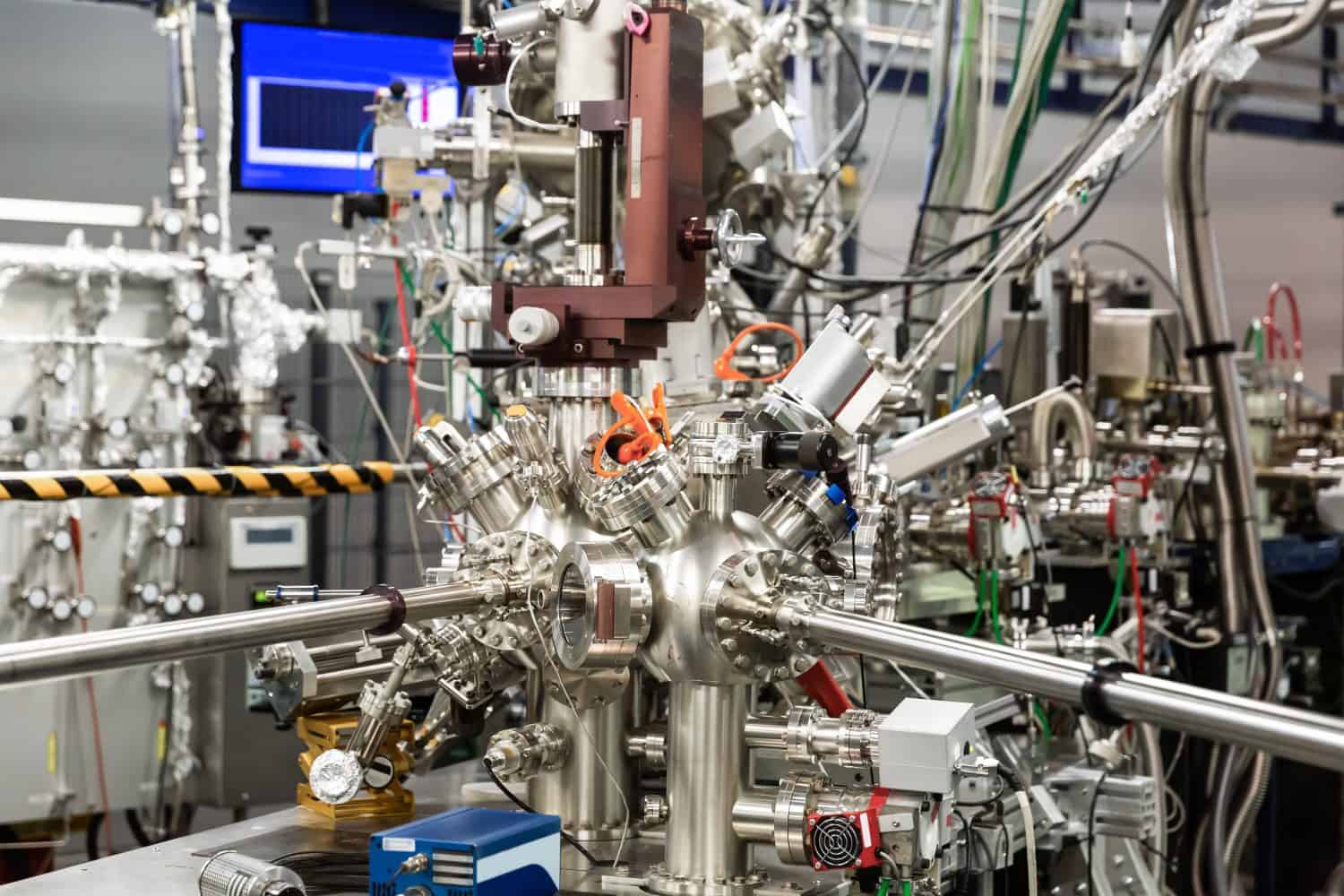
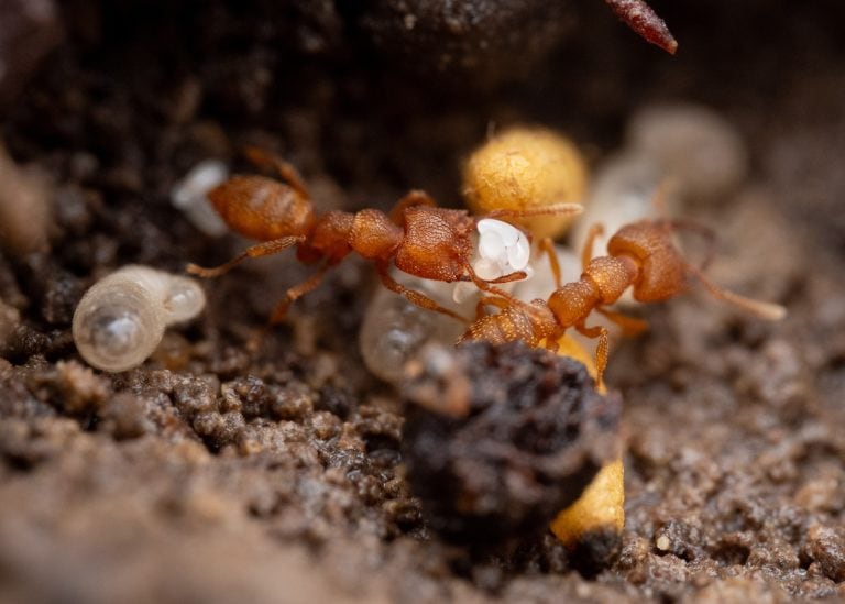
At the Karlsruhe Institute of Technology, a synchrotron particle accelerator produces extremely bright X-ray beams capable of scanning insect specimens in seconds instead of hours.
©BearFotos/Shutterstock.com
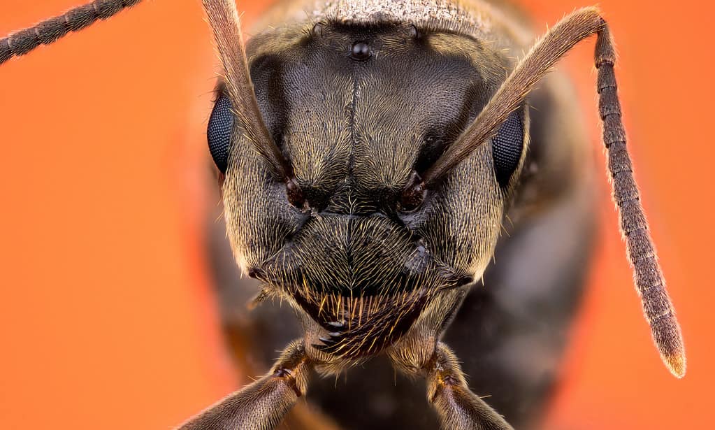
A Detailed Look Inside an Ant
The scans reveal muscles that power an ant’s legs and mandibles. They show the layout of the nervous system, including nerve cords running through the body. Researchers can also examine the digestive tract, glands, and reproductive organs.
In some species, the models clearly show the internal structure of the stinger and venom apparatus. In others, they highlight the thick armor that forms the insect’s exoskeleton.
These anatomical features are critical for scientists studying evolution and functional biology. Small differences in structure can reveal how species adapted to different environments, diets, or social behaviors.
Traditionally, examining such details often required physically dissecting specimens, which destroyed the sample, limiting how many researchers can study it. Digital models not only solved that problem, but now the same scan can be shared with scientists around the world.
The researchers behind Antscan describe their goal as creating a “living library” of organisms. Instead of preserving species only as physical museum specimens, they want to build massive digital collections that anyone can explore.
Early Discoveries from the Antscan Data
The Antscan database is already helping scientists answer new research questions.
In a separate study published in Science Advances in late 2025, researchers used Antscan models to investigate a fascinating evolutionary trade-off in ant colonies. The study examined the thickness of the cuticle, which is the hardened outer layer that forms an ant’s exoskeleton. Building thick armor requires significant nutrients, including nitrogen and minerals. That means producing heavily armored workers can be costly for a colony.
By analyzing hundreds of species, the researchers found a strong negative relationship between cuticle thickness and colony size. Species with thicker exoskeletons tended to have smaller colonies, while species with thinner armor often formed much larger societies.
Basically, some ant species appear to invest in stronger individual soldiers with smaller armies, while others invest in weaker soldiers but with far larger armies.
Before Antscan, measuring cuticle volume across hundreds of species would have been extremely difficult. The digital models made those measurements far easier to perform at large scale.
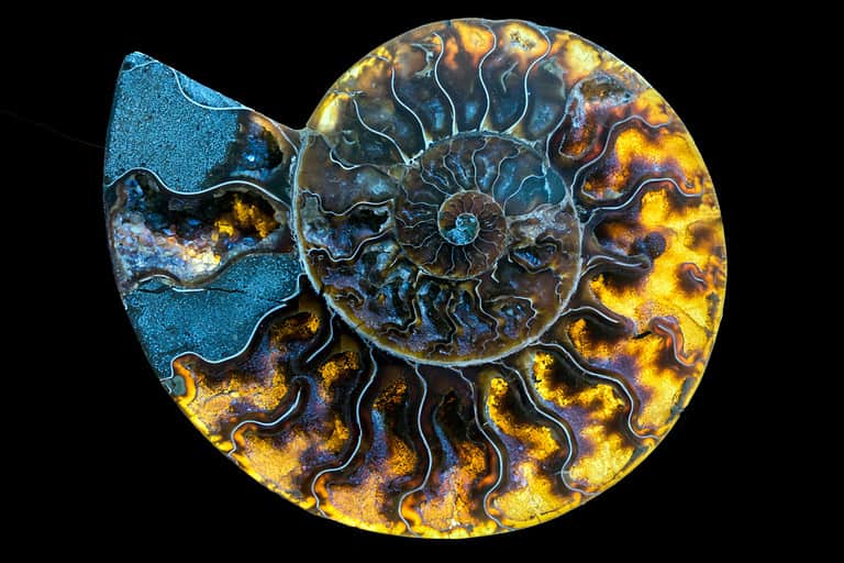
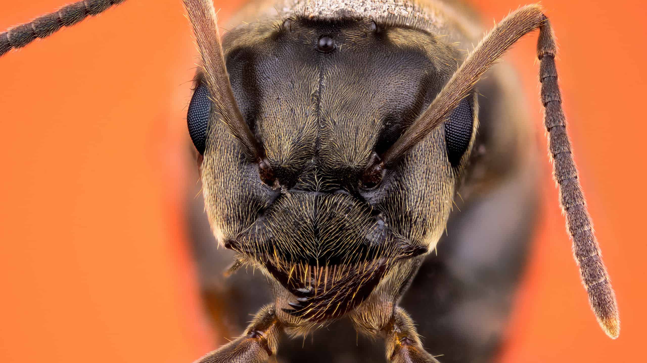
Because the models capture real anatomy in high resolution, they could be used not only in research but also in education, virtual reality, and film animation.
©iStock.com/stockfotocz
From Science Labs to Movie Studios to Classrooms
While Antscan is primarily a scientific project, its applications go far beyond academic research.
High-quality 3D animal models are in constant demand in fields such as animation, game design, and visual effects. Creating realistic insect models for movies or documentaries often take artists weeks or months. Because Antscan models capture real anatomy at extremely high resolution, they could serve as accurate reference material or even direct assets for digital production.
Virtual reality developers might also use the models to create immersive educational experiences. Imagine shrinking down to explore the inside of an ant colony and examine the anatomy of its inhabitants in real time.
Educators could use the same models to bring insect biology to life for students who might never see such detail in a traditional classroom.
Beyond Ants
Antscan represents more than just a new ant database. It demonstrates a powerful workflow that could be applied to countless other organisms.
Museums around the world house billions of preserved specimens, many of which have never been studied in detail. Scanning systems like the one used at Karlsruhe could rapidly digitize these collections.
If similar techniques were applied to beetles, spiders, fish, birds, or even plant structures, scientists could begin building comprehensive digital archives of Earth’s biodiversity.
AI would then help analyze and organize those enormous datasets. Machine learning tools could identify anatomical patterns, classify species, and even detect organisms from field photographs. Such capabilities could dramatically accelerate research in ecology, evolution, and conservation.
Earth’s biodiversity is still far from fully understood. Scientists estimate that millions of species remain undiscovered, and many known species have been studied only superficially. Projects like Antscan offer a glimpse of how technology may change that.
By combining synchrotron X-ray scanning, robotics, and artificial intelligence, researchers were able to scan thousands of ant specimens in days rather than years. The resulting 3D models reveal anatomical details down to muscles, nerves, digestive organs, and stingers.
Even better, those models are freely available to anyone who wants to explore them!
As digital libraries of organisms continue to grow, scientists may soon be able to study life on Earth with an unprecedented level of detail. From evolutionary research to classroom education to cinematic animation, the possibilities feel endless.
For now, Antscan is focused on ants. But its real achievement might be showing how the entire tree of life could someday be digitized, shared, and explored by anyone with an internet connection.