
Quick Take
- Sharks' teeth and fins get all the credit, but their secret speed weapon is hiding somewhere far less obvious. See how vertebrae were studied →
- The same design that makes a great white unstoppable would make a sand tiger shark nearly useless in its own habitat. See species-specific trade-offs →
- This shark spine research may matter more to engineers than to biologists, and the reason why is genuinely surprising. Discover the engineering angle →
When we think of sharks, our minds go to their razor-sharp teeth and signature fins, poking out of the water and warning of potential danger. However, sharks’ status as apex predators is as much due to their spines as to their teeth and fins. With skeletons made of cartilage that has evolved over 400 million years, sharks have gained the dexterity and strength necessary for impressive underwater movements. A new study by researchers at Florida Atlantic University and NOAA Fisheries reveals how hidden designs within shark spines give them the ability to move with endurance, power, and grace.
Scientists have long studied the dynamics of shark musculature and how they produce different swimming styles depending on the species. However, less was known about the structural integrity of shark vertebrae until now. Researchers discovered that complex mineralized structures within shark vertebrae transform rigid spines into powerful, flexible biomechanical systems. Let’s explore this new study and learn how structures hundreds of millions of years in the making have helped sharks survive and thrive in the shifting currents of the world’s oceans.
Uncovering Adaptations
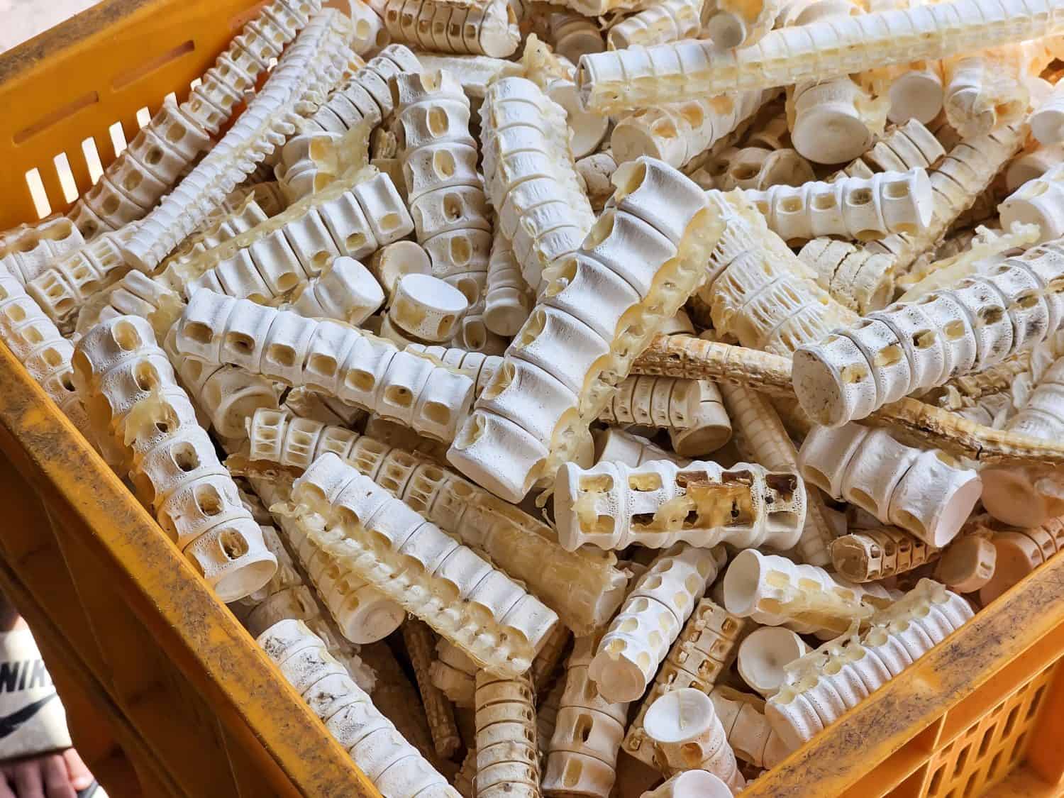
Researchers discovered that the cartilaginous spines of sharks contain specialized mineral structures.
©Howe Hoi/Shutterstock.com
Earlier this month, researchers from Florida Atlantic University and collaborators from NOAA Fisheries published a study in the Journal of Anatomy. They examined the specific structures found in six species of shark vertebrae. This revealed just how well-engineered these systems are for swimming efficiency, flexibility, and strength.
To accomplish this, the research team collected samples from different regions of each shark’s spine and analyzed them using a high-resolution micro-computed tomography system. This produced complete three-dimensional images that allowed for further examination without damaging real vertebrae. Researchers compared each vertebra section and mapped a network of mineralized plates and branching structures. These mappings were compared with the swimming speeds and physiological forms of each shark species: the great white, shortfin mako, porbeagle, common thresher, sand tiger, and basking shark.
As Jamie Knaub explained to EurekAlert, this imaging process gave researchers a glimpse into a hidden world. It also showed how hundreds of millions of years of evolution have made sharks into spectacular swimmers. Knaub, the study’s first author, a research specialist at FAU Laboratory Schools, and a Ph.D. candidate in the FAU Department of Biology, observed that the internal architecture varied among species. He said, “By examining species with varied swimming strategies, we found that the shark spine is not a one-size-fits-all design. Instead, its internal architecture reflects the unique demands of each species’ movement.”
The Findings
One pattern researchers found across the different species was that the middle of shark spines consistently had the largest vertebrae. However, it was the structures within these sections that surprised the researchers the most.
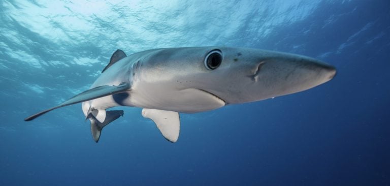
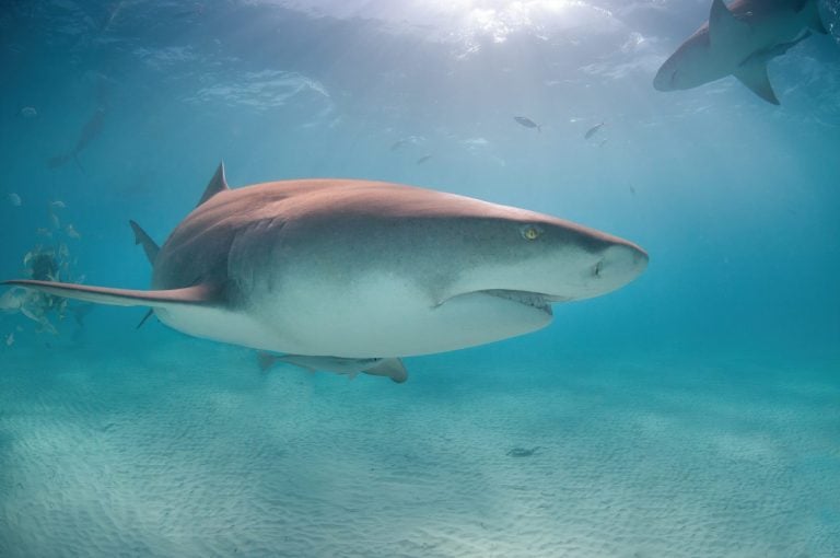
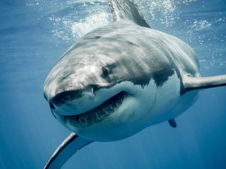
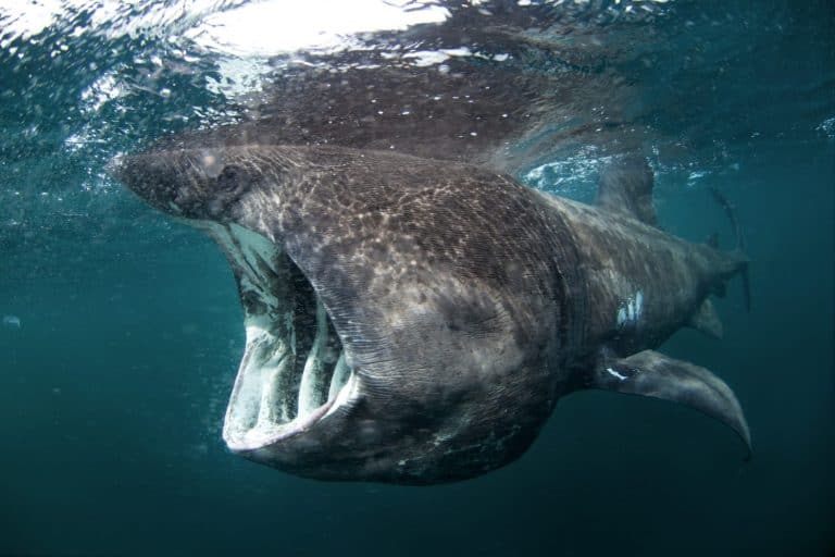
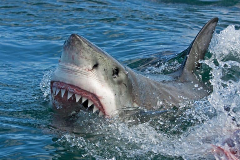
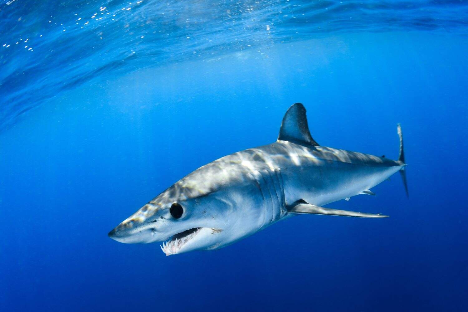
Great white sharks, shortfin makos, and porbeagles are the fastest swimmers among the shark species studied. Each had internal mineralized structures within their vertebrae that helped transfer energy from their bodies to their tails. This more rigid vertebral column effectively propelled great whites and makos through the water at remarkable speeds. Other species studied, like sand tiger sharks, had vertebrae that allowed for more flexibility. This structure enabled slower but more agile movement in complex marine environments. Meanwhile, common thresher sharks had the highest density of mineralized plates and branching structures in their vertebrae, enabling their spines to withstand the force of powerful tail strikes used to stun prey. Basking sharks, being one of the slowest shark species, had reduced mineralization in their spines.
The mineralized structures in shark vertebrae reflect the specific swimming needs of each species. These structures were densest near the tail, particularly in fast-moving shark species. This rigid region near the tail enabled makos and great whites to efficiently transfer energy with each tail stroke. As Marianne E. Porter, Ph.D., senior author and a professor at the FAU Department of Biological Sciences, explained, these structures represent evolution in action. She told EurekAlert, “Nature has spent hundreds of millions of years refining these designs. What we see is an elegant example of biomechanics in action, where each species has evolved a vertebral column precisely tuned to the way it swims. The shark spine isn’t simply flexible—it’s optimized to balance strength, stiffness, and motion in ways that maximize performance.
Broader Implications

A better understanding of shark spines could inspire new technologies optimized for both strength and speed.
©TongTa25/Shutterstock.com
The study shows that closely related shark species, like great whites and shortfin makos, have noticeably similar vertebral designs. In contrast, more distantly related sharks possess very different spine structures. The study was published in the Journal of Anatomy, but the findings may have as many implications for engineering as for biology. The elegant, efficient designs of shark vertebrae could inspire new materials and innovations that are optimized for both strength and agility simultaneously.
Tricia L. Meredith, Ph.D., co-author and director of research for FAU’s on-site lab schools, A.D. Henderson University School and FAU High School, and an assistant research professor in FAU’s College of Education, echoed this view. She told EurekAlert, “These imaging capabilities allow us to move beyond simply describing anatomy to understanding how internal structures function mechanically, opening the door to new discoveries in comparative biology and biomimetic design.”