
Quick Take
- A new study shows that zebrafish larvae use neurons and hormones to brighten and darken themselves
- This pathway acts like a “dimmer switch” as opposed to all-or-nothing
- Learning about this can help experts better understand other animal camouflage processes.
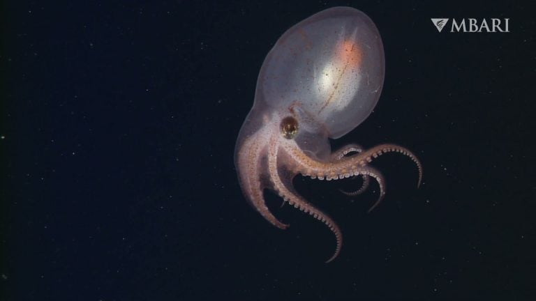
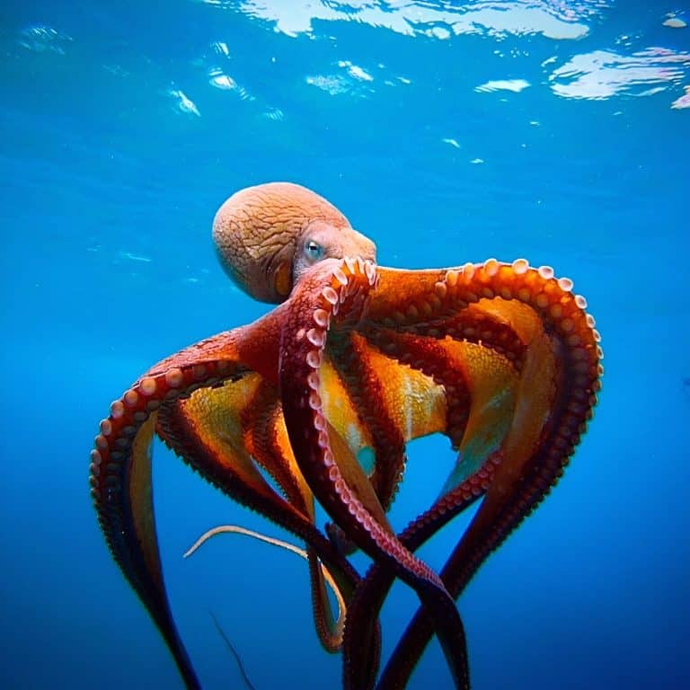
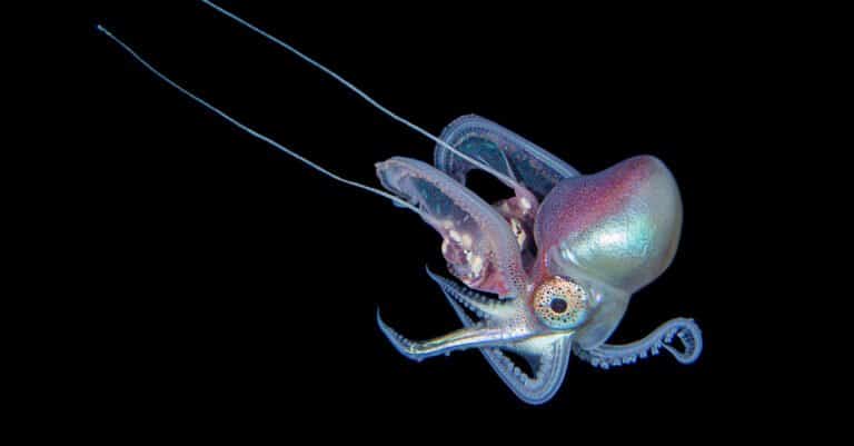
If you’ve ever watched an animal seem to “melt” into its surroundings, you’ve seen background adaptation in action. An octopus darkens to match the rocks around it. A chameleon changes color to blend into its environment. It’s one of nature’s sneakiest defenses: match the brightness of the environment, become harder to spot, and live to swim another day.
Zebrafish larvae—tiny, see-through fish often used in biology labs—do this too. When they move into brighter areas, they gradually fade lighter over the course of minutes. For a predator looking down from above, that shift could mean the difference between a quick snack and a missed target.
Scientists have now gone beyond identifying what happens and have mapped out how it happens. A research team at the Max Planck Institute for Biological Intelligence traced the whole chain of command that links light detection in the eye to hormone signals in the brain and, finally, to pigment movement in skin cells. Their findings were published in Current Biology.
How Does the Camouflage System Work?
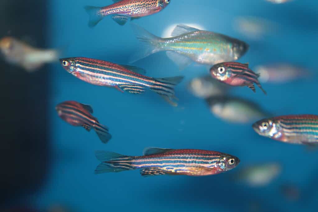
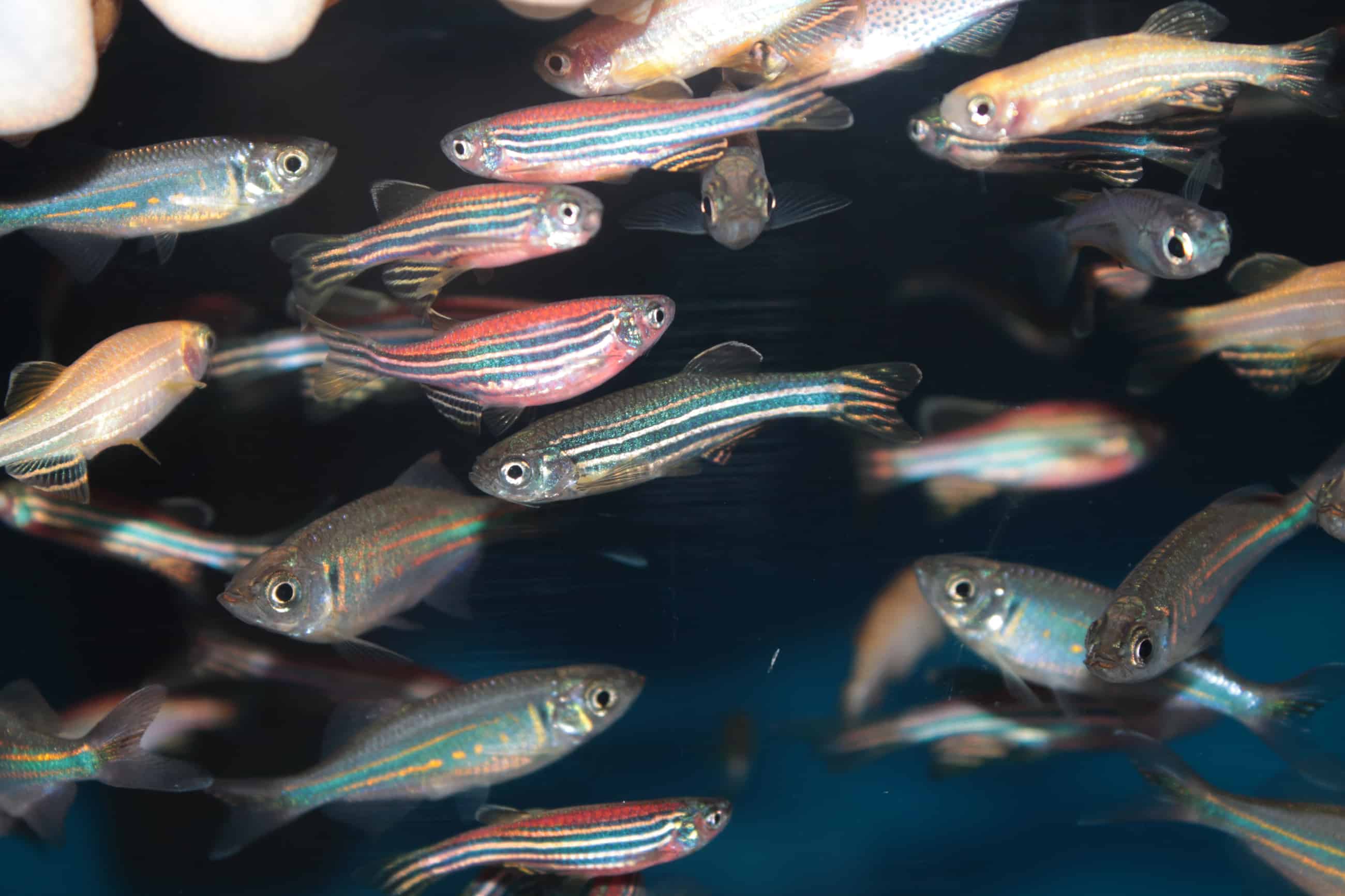
Zebrafish (Danio rerio) is a tropical aquarium fish
©kazakovmaksim/iStock via Getty Images
The zebrafish larva’s shifting color isn’t driven by a single “turn pale” button in the skin. Instead, it’s controlled by specialized pigment cells called melanophores, each packed with tiny bundles of melanin, the same pigment family that colors human skin and hair. The way these pigment particles are arranged determines the fish’s appearance.
When the melanin gathers into tight clusters, the young fish appears lighter overall. When it spreads throughout the cell, the animal looks darker. In that sense, camouflage is less like painting a new coat and more like directing traffic inside living cells, constantly repositioning pigment to match the brightness of the world outside.
What makes the new research especially striking is that the process doesn’t begin in the skin at all; it actually starts in the eye. The study identifies retinal nerve cells as the key starters of the chain reaction. These are neurons that carry information from the retina into the brain, and here they play a central role in translating ambient light into a body-wide response.
“Retinal ganglion cells in the eye detect light and transmit this signal in the form of neuronal activity to neurons in the brain’s hypothalamus. These, in turn, produce a hormone and secrete it into the bloodstream, causing the fish to lighten. At the same time, again acting on retinal ganglion cells, light suppresses a different hormone in a different population of neurons that darkens the skin,” Dr. Krasimir Slanchev, a senior scientist at the Max Planck Institute for Biological Intelligence and the paper’s first author, said in a statement.
This isn’t a simple, one-way order that constantly pushes the skin toward paleness. Instead, the chemical pathway is designed to balance in both directions depending on lighting conditions, creating a controlled shift rather than an all-or-nothing flip. Slanchev captured that push-pull design with an everyday comparison: “The system works like a dimmer switch with two controls working in opposite directions.”
In other words, the brightness doesn’t just turn up a pathway that lightens the fish; it also turns down a separate pathway that would otherwise darken it. By coupling activation with suppression, the animal can fine-tune its appearance as the environment changes.
That “dimmer switch” analogy fits because the fish’s hormones act as long-range messengers between brain and skin. In the study, the researchers followed how light-driven signals shift opposing hormonal influences, one that promotes melanin clustering and a lighter look, and another that promotes pigment dispersal and a darker look. The result of those hormone adjustments is a change in how melanophores arrange their melanin packets, which is what the predator—or the microscope—ultimately sees.
How to See the Molecular Pathways
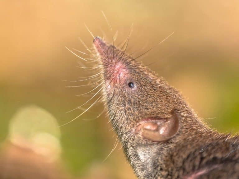
Much of the camouflage process for zebrafish actually starts in the nerve cells in its eye.
©iStock.com/whitehoune
Zebrafish larvae are unusually useful for answering this kind of question because they’re transparent and genetically accessible. The researchers used fluorescent labels to visualize connections, genetic tools to target specific cells, and experiments that removed or silenced certain components to see what broke.
By combining these techniques, the researchers were able to trace the entire pathway from the retina, through the brain and hormones, to the skin’s response—going beyond the previously understood step of the retina simply sensing light.
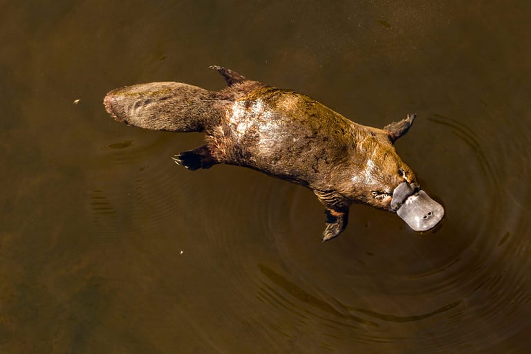
How Does This Help Us Better Understand Animal Camouflage?

Understanding camouflage processes can help us with conservation efforts
©Steve Allen/Shutterstock.com
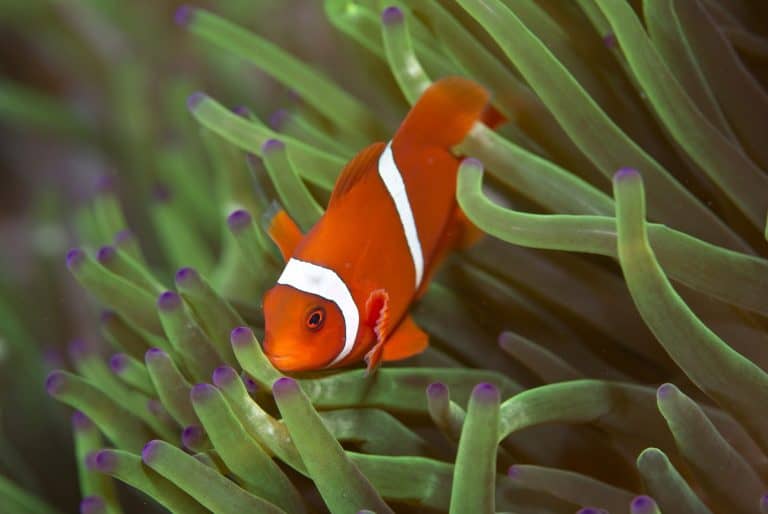
Color change isn’t only about hiding. Across animals, it influences courtship, hunting, and social signaling too. As department director, Herwig Baier, said in the press release, “Color change is critical for how some animals evade predators, find mates, hunt, and navigate their lives.”
Baier also points to the broader biological question behind the discovery: once you can identify the precise cells and connections underlying a behavior, you can start asking how evolution shaped (or removed) those circuits across species. “This work opens broader questions about how sensory information transforms into hormonal signals and how this ancient vertebrate response was lost when mammals evolved—giving us a window into a survival mechanism our ancestors left behind,” he said.